ORIGINAL ARTICLE
https://doi.org/10.47811/bhj.223
Antimicrobial Susceptibility Pattern and Proportion of Multidrug-Resistant Organisms among Patients with Bloodstream Infections at Eastern Regional Referral Hospital, Bhutan: A Retrospective Descriptive Study
Sanga Tenzin1, Ugyen Rinzin2, Pelden Wangchuk3, Chimi Wangmo4, Nima Dorji4, Ugyen Chenzom1, Tsheten Wangchuk5, Yeshey Dorjey6
1Department of Pharmacy, Eastern Regional Referral Hospital, Mongar, Bhutan 2Department of Emergency Medicine, Eastern Regional Referral Hospital, Mongar, Bhutan
3Office of the Medical Superintendent, Eastern Regional Referral Hospital, Mongar, Bhutan
4Department of Pathology and Laboratory Medicine, Eastern Regional Referral Hospital, Mongar, Bhutan
5Department of Surgery, Eastern Regional Referral Hospital, Mongar, Bhutan 6Department of Obstetrics and Gynecology, Eastern Regional Referral Hospital, Mongar, Bhutan
Corresponding author:
Sanga Tenzin
sangatenzin3@gmail.com
ABSTRACT
Introduction: Growing antimicrobial resistance to commonly used antimicrobial exacerbates global burden of the bloodstream infection (BSIs). Understanding the local antimicrobial susceptibility pattern is crucial for effective antimicrobial stewardship. This study evaluated the antimicrobial susceptibility pattern and proportion of multidrug-resistant organisms (MDROs) among blood culture bacterial isolates at Eastern Regional Referral Hospital. Methods: The retrospective study reviewed blood culture records of 2025. A total of 79 positive blood cultures from 62 unique patients were included. Antimicrobial susceptibility testing was performed using standard microbiological methods and interpreted according to the Clinical and Laboratory Standards Institute (CLSI) guidelines. Data were collected in REDCap and analyzed using SPSS version 32 and R version 4.6.0. Results: Of 981 blood cultures performed in 2025, 8.1% showed positive culture. Most cases occurred in patients over 40 years (74.7%), with a female predominance (53.2%). Sepsis was the predominant clinical diagnosis (78.5%), and 70.9% had comorbidities. Gram-negative organisms accounted for 78.5% of isolates led by E. coli (45.2%), which exhibited high susceptibility to amikacin (86.7%). Staphylococcus aureus (82.4%) was the main Gram-positive isolate demonstrating complete susceptibility to cloxacillin (100%). Overall, 23.0% (17/74) of the isolates were MDROs. For carbapenem resistance, 35.7% (5/14) of tested isolates were identified as CRO, representing 6.3% (5/79) of the total cohort. Conclusion: Gram-negative organisms, particularly, E. coli was the main causative pathogen. The findings provide vital insights into the local susceptibility patterns to guide antimicrobial stewardship programs.
Keywords: Antimicrobial resistance; Antimicrobial susceptibility; Blood culture; Escherichia coli; Gram-negative bacteria; Gram-positive bacteria; Multidrug-resistance; Sepsis
INTRODUCTION
BSI is one of the life-threatening medical emergencies associated with high rates of morbidity, clinical complications and mortality worldwide1. BSI is characterized by the presence of viable microorganisms in the bloodstream, usually detected through positive blood culture, in association with clinical signs of infection and systemic inflammatory response2. BSI may occur as a complication of various clinical conditions including pneumonia and deep tissue infections such as septic arthritis 3,4. Furthermore, BSI can cause acute infection and trigger sepsis a life-threatening host immune response to an infection which causes severe organ dysfunction 5,6.
Blood culture remains the "gold standard" for diagnosis of BSI allowing identification of the pathogens and antimicrobial susceptibility testing 7. Blood culture is indicated for conditions with a strong likelihood of BSI such as septic shock, endovascular infections, pneumonia, septic joints and meningitis 8.
Studies from different regions have shown varying blood culture positivity rates with commonly isolated pathogens including Gram-positive bacteria such as Staphylococcus aureus and Gram-negative bacteria such as E. coli, Klebsiella pneumoniae and Pseudomonas aeruginosa 9,10.
In the Southeast Asia region including India, Nepal, Pakistan and Bangladesh, the rate of positive blood culture generally ranges from approximately 12% to 30% 11-14. Reflecting the global trends, the predominant pathogens in the region include Staphylococcus aureus, E. coli, Klebsiella pneumoniae and Pseudomonas aeruginosa 12,13,15.
Beyond the high burden of the infection, the global expansion of antimicrobial resistance (AMR) among predominant pathogens has further complicated the clinical management of BSI with a disproportionate burden in low- and middle-income countries (LMICs) 16. Evidence from Nepal documented an alarming rise in multidrug-resistant organism (MDRO) from 20% to 43% and carbapenem-resistant Enterobacterales from near zero to 65% over the years 13. In India, high resistance to third generation cephalosporins in E. coli and Klebsiella pneumoniae, with additional carbapenem-resistance ranging from 20% to 65% has been documented across multiple centers 17.
Within the national healthcare framework of Bhutan, regional referral hospitals serve as the main medical hub managing complex infections. The Eastern Regional Referral Hospital (ERRH) serves as an essential tertiary care center for the eastern region of the country, receiving critically ill patients across departments such as medical, surgical and emergency. Although Bhutan has national antibiotic guidelines and a relatively regulated antibiotic supply system, regional variations in pathogen prevalence and antibiotic resistance can vary significantly due to localized antibiotic prescribing patterns and varying patient demographics. Therefore, locally generated surveillance data are essential to inform empirical treatment decisions and support effective antimicrobial stewardship interventions. However, comprehensive data regarding the bacterial profile and antibiotic susceptibility patterns in this setting remain limited. The gap in the surveillance data may compromise the precision of empirical therapy and hinder optimization of antibiotic stewardship programs. To address these challenges, this study aimed to evaluate the antibiotic susceptibility patterns and the proportion of MDROs among blood culture bacterial isolates from patients with BSI at ERRH.
MATERIALS AND METHODS
Study design
A retrospective descriptive study was conducted at ERRH, Mongar, Bhutan.
Study setting
The study was conducted at a 150-bed hospital, which serves as an essential referral center for eastern region of Bhutan. The hospital provides a wide range of specialized clinical services through various departments including Medicine, Emergency Medicine, General Surgery, Obstetrics & Gynecology, Pediatrics, Orthopedics, Ophthalmology, and Otorhinolaryngology.Laboratory diagnostic support is provided by the Department of Pathology & Laboratory Medicine. Diagnostic services include hematology, biochemistry, blood banking, anatomical pathology and microbiology services. The microbiology laboratory provides antibiotic susceptibility testing for various clinical specimens including blood.
Study period
Medical records documented at the Microbiology Lab and medical record unit from 1st January to 31st December 2025 were retrospectively reviewed and analyzed from 1st January to 31st January 2026 after receiving an ethical clearance waiver.
Study population
All 981 blood culture specimens submitted to the Microbiology Laboratory in the year 2025 were evaluated. The unit of analysis was the blood culture isolate recovered from each positive blood culture episode. A total of 79 positive blood culture episodes were recovered from 62 unique patients. Each positive blood culture isolate was analyzed independently. Consequently, 17 isolates recovered from the same patient on different dates were retained because they represented separate blood culture episodes.
Inclusion criteria
All positive blood culture episodes, including repeat positive culture episodes from the same patient on different dates, were included in the study.
Exclusion criteria
Those blood samples with negative growth on culture and record with missing and incomplete information were excluded from the study. In addition, coagulase-negative staphylococci (n = 77) were also excluded since no susceptibility test was performed as per the institutional protocol due to their high likelihood of representing skin contaminants.
Sample size and sampling method
Records of all blood culture specimens processed at the microbiology laboratory during 2025 were reviewed, and all case fulfilling inclusion criteria were included for the study, and a separate sample size was not determined. A consecutive sampling method was employed for data collection.
Study procedure
Laboratory register maintained at microbiology laboratory and medical record of the patients were retrospectively reviewed and relevant variables were extracted using a structured proforma developed in REDCap. Demographic characteristics (age, gender), departments, clinical characteristics, previous antibiotic exposure, microbial isolates of blood samples, and patterns of antimicrobial susceptibility (susceptible, intermediate and resistant), multidrug-resistant organisms, and carbapenem-resistant organisms were all collected.
Microbiological methods
Blood samples were collected according to the standard operating procedures of the Microbiology Laboratory. For adults 8-10 ml of blood were collected and for pediatrics 2-5 ml were collected from the median cubital vein for each blood culture bottle. One blood culture set from venipuncture site was collected before antimicrobial therapy. Blood cultures were incubated at 37 ℃ using BACTECTM (Becton, Dickinson Microbiology Systems) and BACT/ALERT® system for a minimum of 5 days. Both the BACTECTM and BACT/ALERT® automated blood culture systems were used in the microbiology laboratory. Blood culture specimens were processed using the system compatible with the blood culture bottles available at the time of specimen collection. Thus, specimen allocation was based solely on bottle compatibility with the respective instrument. Positive blood culture bottles detected by the BACTECTM and BACT/ALERT® blood culture system were sub-cultured onto MacConkey agar and 5% sheep blood agar plates. The inoculated plates were incubated at 35-37℃ for 24-48 hours under appropriate atmospheric conditions, after which bacterial growth was examined and isolates were processed for identification and antimicrobial susceptibility testing. Bacterial isolates were identified using conventional microbiological methods, including assessment of colony morphology, gram staining, and standard biochemical tests. Isolates requiring further confirmation were identified using the Analytical Profile Index identification system. Antimicrobial susceptibility testing was performed by Kirby Bauer disk diffusion method and interpreted according to the CLSI, Performance Standards for Antimicrobial Susceptibility Testing, M100, 33rd edition 2023. Antibiotic susceptibility testing was performed tier-wise - testing was performed for first tier, and if detected intermediate or resistant to first tier, a subsequent tier antibiotic was tested. Consequently, susceptibility percentages reported for second-tier agents represent conditional probabilities and reflect potential selection bias, as these agents were tested primarily on isolates enriched for resistance. Cefoxitin disk testing was used as a surrogate for methicillin or oxacillin resistance and reported as cloxacillin susceptibility 18. Quality control was performed using standard reference strains, including Escherichia coli ATCC 25922, Staphylococcus aureus ATCC 25923, and Pseudomonas aeruginosa ATCC 27853.
Operational definitions
Multidrug-resistant organism - It is defined as "acquired non-susceptibility to at least one agent in three or more antimicrobial categories." An isolate was classified as a multidrug-resistant organism when it demonstrated acquired non-susceptibility (i.e., intermediate or resistant interpretation) to at least one antimicrobial agent in three or more antimicrobial classes, according to the international consensus definition 19. To ensure calculation reproducibility, antimicrobial classes were defined separately by organism group, excluding intrinsic resistance and clinical non-recommended combinations. Isolates were considered evaluable for MDRO classification only if susceptibility results were available for at least three eligible antimicrobial classes. These criteria were applied to E. coli, Klebsiella pneumoniae, Staphylococcus aureus, Acinetobacter spp., Enterobacter sp., Citrobacter sp., and 16 Pseudomonas aeruginosa isolates. Conversely, 2 Pseudomonas aeruginosa and 3 Streptococcus sp. isolates had fewer than three tested classes; consequently, they were classified as non-evaluable and excluded from the MDRO denominator. Overall, 74 of the 79 total isolates met the criteria for complete testing and were included in the MDRO analysis.
Carbapenem-resistant organism - An isolate was classified as a CRO when it demonstrated resistance to at least one tested carbapenem agent (imipenem or meropenem) according to the CLSI antimicrobial susceptibility testing interpretive criteria. CRO classification was performed at the isolate level based on the susceptibility results of each blood culture isolates 20.
WHO AWaRe classification of antibiotics - To assist in the development of tools for antibiotics stewardship program and to reduce antibiotic resistance, AWaRe classification of antibiotics (access, watch, and reserve) was developed by World Health Organization 21.
Access group - includes antibiotics that have activity against a wide range of commonly encountered susceptible pathogens while also showing lower resistance potential than antibiotics in the other groups. Access group of antibiotics are recommended as first or second line of empirical treatment 21.
Watch group - includes antibiotics that have higher resistance potential and includes most of the highest priority agents among the critically important antimicrobials. These antibiotics are recommended as the first or second line of empiric treatment of a specific infection 21.
Reserve group- includes antibiotics that should be reserved for treatment of confirmed or suspected infections due to MDRO. These antibiotics should be treated as "last resort" options 21.
Data management and confidentiality
A unique identifier was allocated to every isolate enrolled in the study, and the investigators carefully entered data in REDCap and the data were double checked for duplicate entries. Supervision of the data collection and a thorough review of form completeness were conducted by the principal investigator. All eligible positive blood culture isolates had complete clinical and laboratory documentation, with zero records excluded for missing data or incomplete entry. A clear distinction was maintained between missing records and unperformed susceptibility tests: variations in testing denominators (N) across antimicrobial agents were strictly driven by the laboratory's selective, tier-wise testing protocol rather than missing data records. Confidentiality was maintained by keeping the patient's data in a secure folder locked with a password. The questionnaire did not include any identifying data of the patients.
Data analysis
Data were captured using REDCap (Research Electronic Data Capture). The dataset was then exported and analyzed using SPSS (Version 32) (IBM Corp., Armonk, NY, USA) and R (Version 4.6.0). Descriptive analysis was performed and data were presented as frequencies and percentages, and a heatmap was used to present the antimicrobial susceptibility.
RESULTS
A total of 981 blood cultures were performed in 2025. Excluding 77 coagulase-negative staphylococci isolates deemed contaminants per institutional protocol, 8.1% (n = 79) yielded positive cultures (62 unique patients, and 17 were repeated blood cultures performed on different dates).
Demographic, Microbiological and Clinical Characteristics
The positive blood culture was predominant among older patients, with nearly three-quarters of them aged over 40 years (74.7%, n = 59) and over half were female (53.2 %, n = 42). More than three-quarters of the patients were clinically diagnosed with sepsis (78.5%, n = 62), and the majority (70.9%) had underlying comorbidities.
Microbiological analysis revealed a predominance of Gram-negative organisms (78.5%, n = 62), of which E. coli (45.2%, 28/62) was the most isolated, followed by Pseudomonas aeruginosa (29.0%, 18/62). Meanwhile, Staphylococcus aureus was the most frequently isolated Gram-positive organism (82.4%, 14/17). Detailed data is presented in Table 1.
Table 1: Demographic, Clinical, and Microbiological Characteristics of patients with bloodstream infection at the Eastern Regional Referral Hospital, Mongar, Bhutan, 2025 (n = 79).
|
Characteristics |
Frequency |
Percent |
|
|
Age in years |
|
|
|
|
|
<20 years |
13 |
16.5 |
|
|
20-40 years |
7 |
8.9 |
|
|
>40 years |
59 |
74.7 |
|
Sex |
|
|
|
|
|
Female |
42 |
53.2 |
|
|
Male |
37 |
46.8 |
|
Departments |
|
|
|
|
|
Emergency |
13 |
16.5 |
|
|
Medicine |
34 |
43.0 |
|
|
Orthopedics |
9 |
11.4 |
|
|
Pediatrics |
6 |
7.6 |
|
|
Surgery |
17 |
21.5 |
|
Clinical Diagnosis |
|
|
|
|
|
Sepsis |
62 |
78.5 |
|
|
Pneumonia |
9 |
11.4 |
|
|
Joint Infections |
8 |
10.1 |
|
Comorbidities |
|
|
|
|
|
Present |
56 |
70.9 |
|
|
Absent |
23 |
29.1 |
|
Gram-staining |
|
|
|
|
|
Positive |
17 |
21.5 |
|
|
Negative |
62 |
78.5 |
|
Gram positive |
|
|
|
|
|
Staphylococcus aureus |
14 |
82.4 |
|
|
Streptococcus spp. |
3 |
17.6 |
|
Gram negative |
|
|
|
|
|
Acinetobacter spp. |
2 |
3.2 |
|
|
Citrobacter sp. |
1 |
1.6 |
|
|
Enterobacter sp. |
1 |
1.6 |
|
|
E. coli |
28 |
45.2 |
|
|
Klebsiella pneumoniae |
12 |
19.4 |
|
|
Pseudomonas aeruginosa |
18 |
29.0 |
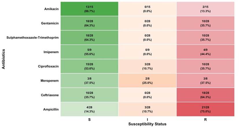
Antimicrobial susceptibility of organisms
E. coli isolates demonstrated the highest susceptibility to amikacin (86.7%), followed by gentamicin (64.3%) and sulphamethoxazole-trimethoprim(64.3%).Pseudomonas aeruginosa showed complete sensitivity to amikacin (100%) and high sensitivity to sulphamethoxazole-trimethoprim (94.4%) and gentamicin (88.9%). Meanwhile, Staphylococcus aureus exhibited complete sensitivity to cloxacillin (100%), alongside high sensitivity to erythromycin (92.9%) and sulphamethoxazole-trimethoprim (85.7%). Other isolates demonstrated variable susceptibility as detailed in Table 2
Table 2: Antimicrobial Susceptibility Pattern of Organisms isolated from patients with Bloodstream Infections at the Eastern Regional Referral Hospital, Mongar, Bhutan, 2025 (n = 79)
|
Antibiotics
|
E.coli, n/N (%) |
PA, n/N (%) |
KP, n/N (%) |
Acin. spp. n/N (%) |
Citro. sp. n/N (%) |
Entero. sp. n/N (%) |
Staph aureus, n/N (%) |
Strep spp. n/N (%) |
|
|
Ampicillin |
|||||||||
|
|
S |
4/28 (14.3) |
X |
X |
X |
0/1 (0.0) |
X |
X |
X |
|
|
I |
3/28 (10.7) |
X |
X |
X |
0/1 (0.0) |
X |
X |
X |
|
|
R |
21/28 (75.0) |
X |
X |
X |
1/1 (100.0) |
X |
X |
X |
|
Penicillin G |
|||||||||
|
|
S |
X |
X |
X |
X |
X |
X |
0/9 (0.0) |
3/3 (100.0) |
|
|
I |
X |
X |
X |
X |
X |
X |
0/9 (0.0) |
0/3 (0.0) |
|
|
R |
X |
X |
X |
X |
X |
X |
9/9 (100.0) |
0/3 (0.0) |
|
Cloxacillin |
|||||||||
|
|
S |
X |
X |
X |
X |
X |
X |
14/14 (100.0) |
X |
|
|
I |
X |
X |
X |
X |
X |
X |
0/14 (0.0) |
X |
|
|
R |
X |
X |
X |
X |
X |
X |
0/14 (0.0) |
X |
|
Piperacillin |
|||||||||
|
|
S |
X |
12/15 (80.0) |
0/1 (0.0) |
X |
X |
X |
X |
X |
|
|
I |
X |
0/15 (0.0) |
1/1 (100.0) |
X |
X |
X |
X |
X |
|
|
R |
X |
3/15 (20.0) |
0/1 (0.0) |
X |
X |
X |
X |
X |
|
Ceftazidime |
|||||||||
|
|
S |
X |
11/13 (84.6) |
X |
X |
X |
X |
X |
X |
|
|
I |
X |
1/13 (7.7) |
X |
X |
X |
X |
X |
X |
|
|
R |
X |
1/13 (7.7) |
X |
X |
X |
X |
X |
X |
|
Ceftriaxone |
|||||||||
|
|
S |
10/28 (35.7) |
X |
5/12 (41.7) |
X |
0/1 (0.0) |
0/1 (0.0) |
X |
X |
|
|
I |
0/28 (0.0) |
X |
2/12 (16.7) |
X |
0/1 (0.0) |
0/1 (0.0) |
X |
X |
|
|
R |
18/28 (64.3) |
X |
5/12 (41.7) |
X |
1/1 (100.0) |
1/1 (100.0) |
X |
X |
|
Ciprofloxacin |
|||||||||
|
|
S |
15/28 (53.6) |
17/18 (94.4) |
8/12 (66.7) |
2/2 (100.0) |
1/1 (100.0) |
1/1 (100.0) |
X |
X |
|
|
I |
3/28 (10.7) |
1/18 (5.6) |
2/12 (16.7) |
0/2 (0.0) |
0/1 (0.0) |
0/1 (0.0) |
X |
X |
|
|
R |
10/28 (35.7) |
0/18 (0.0) |
2/12 (16.7) |
0/2 (0.0) |
0/1 (0.0) |
0/1 (0.0) |
X |
X |
|
Doxycycline |
|||||||||
|
|
S |
X |
X |
X |
X |
X |
X |
11/14 (78.6) |
X |
|
|
I |
X |
X |
X |
X |
X |
X |
2/14 (14.3) |
X |
|
|
R |
X |
X |
X |
X |
X |
X |
1/14 (7.1) |
X |
|
Tetracycline |
|||||||||
|
|
S |
X |
X |
X |
X |
X |
X |
7/14 (50.0) |
X |
|
|
I |
X |
X |
X |
X |
X |
X |
3/14 (21.4) |
X |
|
|
R |
X |
X |
X |
X |
X |
X |
4/14 (28.6) |
X |
|
Sulphamethoxazole-Trimethoprim |
|||||||||
|
|
S |
18/28 (64.3) |
X |
6/12 (50.0) |
2/2 (100.0) |
1/1 (100.0) |
1/1 (100.0) |
12/14 (85.7) |
X |
|
|
I |
0/28 (0.0) |
X |
0/12 (0.0) |
0/2 (0.0) |
0/1 (0.0) |
0/1 (0.0) |
0/14 (0.0) |
X |
|
|
R |
10 /28 (35.7) |
X |
6 /12 (50.0) |
0/2 (0.0) |
0/1 (0.0) |
0/1 (0.0) |
2/14 (14.3) |
X |
|
Erythromycin |
|||||||||
|
|
S |
X |
X |
X |
X |
X |
X |
13/14 (92.9) |
X |
|
|
I |
X |
X |
X |
X |
X |
X |
1/14 (7.1) |
X |
|
|
R |
X |
X |
X |
X |
X |
X |
0/14 (0.0) |
X |
|
Gentamicin |
|||||||||
|
|
S |
18/28 (64.3) |
16/18 (88.9) |
10/12 (83.3) |
2/2 (100.0) |
1/1 (100.0) |
0/1 (0.0) |
X |
X |
|
|
I |
0/28 (0.0) |
0/18 (0.0) |
0/12 (0.0) |
0/2 (0.0) |
0/1 (0.0) |
0/1 (0.0) |
X |
X |
|
|
R |
10/28 (35.7) |
2/18 (11.1) |
2/12 (16.7) |
0/2 (0.0) |
0/1 (0.0) |
1/1 (100.0) |
X |
X |
|
Amikacin |
|||||||||
|
|
S |
13/15 (86.7) |
8/8 (100.0) |
6/6 (100.0) |
2/2 (100.0) |
1/1 (100.0) |
X |
X |
X |
|
|
I |
0/15 (0.0) |
0/8 (0.0) |
0/6 (0.0) |
0/2 (0.0) |
0/1 (0.0) |
X |
X |
X |
|
|
R |
2/15 (13.3) |
0/8 (0.0) |
0/6 (0.0) |
0/2 (0.0) |
0/1 (0.0) |
X |
X |
X |
|
Imipenem |
|||||||||
|
|
S |
5/9 (55.6) |
3/5 (60.0) |
X |
X |
X |
X |
X |
X |
|
|
I |
0/9 (0.0) |
2/5 (40.0) |
X |
X |
X |
X |
X |
X |
|
|
R |
4/9 (44.4) |
0/5 (0.0) |
X |
X |
X |
X |
X |
X |
|
Meropenem |
|||||||||
|
|
S |
3/8 (37.5) |
2/3 (66.7) |
X |
X |
X |
X |
X |
X |
|
|
I |
2/8 (25.0) |
0/3 (0.0) |
X |
X |
X |
X |
X |
X |
|
|
R |
3/8 (37.5) |
1/3 (33.3) |
X |
X |
X |
X |
X |
X |
|
Piperacillin/Tazobactam |
|
|
|
|
|
|
|
||
|
|
S |
X |
0/1 (0.0) |
X |
X |
X |
X |
X |
X |
|
|
I |
X |
1/1 (100.0) |
X |
X |
X |
X |
X |
X |
|
|
R |
X |
0/1 (0.0) |
X |
X |
X |
X |
X |
X |
Acin. spp: Acinetobacter species; Citro. sp: Citrobacter species; Entero. sp: Enterobacter species; I: intermediate; KP: Klebsiella pneumoniae; PA: Pseudomonas aeruginosa; R: resistant; S: susceptible; X: test not performed; n/N: 'n' indicates the number of isolates in the S, I, or R category, and 'N' indicates total number of isolates tested for that organism-antibiotic pair.
Note. Second-tier antimicrobial agents were tested selectively following a selective reporting protocol, conditioned on non-susceptibility to first-tier agents. Susceptibility percentages for second-tier agents reflect only this pre-selected subset (e.g., amikacin tested on 15 of 28 E. coli isolates) and are not directly comparable to first-tier susceptibility rates. Cloxacillin susceptibility was determined using cefoxitin disk diffusion testing as a surrogate marker.

Figure 1: Heatmap of antimicrobial susceptibility pattern of E. coli among patients with Bloodstream Infection at the Eastern Regional Referral Hospital, Mongar, Bhutan, 2025
*Note. S = susceptible; I = intermediate; R = resistant. Numbers inside cells represent n/N (%), where 'n' indicates the number of isolates in the S, I, or R category, and 'N' indicates total number of isolates tested for that organism-antibiotic pair. N per antibiotic ranges from 8 to 28 (see Table 2 for exact denominators).
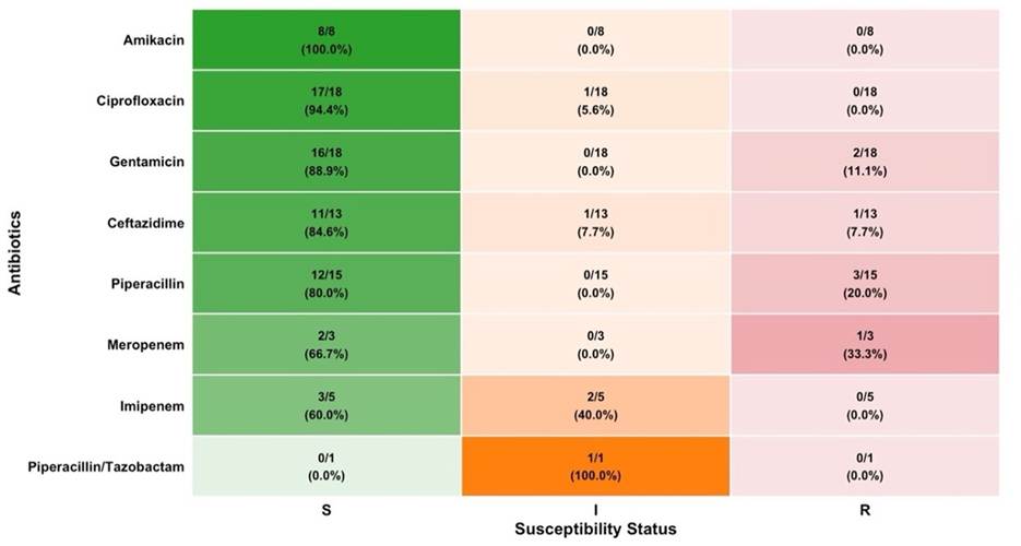
Figure 2: Heatmap of antimicrobial susceptibility pattern of Pseudomonas aeruginosa among patients with Bloodstream Infection at the Eastern Regional Referral Hospital, Mongar, Bhutan, 2025
*Note. S = susceptible; I = intermediate; R = resistant. . Numbers inside cells represent n/N (%), where 'n' indicates the number of isolates in the S, I, or R category, and 'N' indicates total number of isolates tested for that organism-antibiotic pair. N per antibiotic ranges from 1 to 18 (see Table 2 for exact denominators).
Previous antibiotic exposure pattern
Out of 79 blood culture positive episodes with a complete exposure history, over 60% (48/79) had prior antibiotic exposure. Individual episode could involve exposure to more than one antibiotic. Over three-fifth were exposed to doxycycline (62.5%, 30/48) followed by ceftriaxone (37.5%, 18/48) and ampicillin (29.2%, 14/48) before the blood culture collection. The exposure to other antibiotics varied and occurred at lower frequencies as presented in the Figure 3.
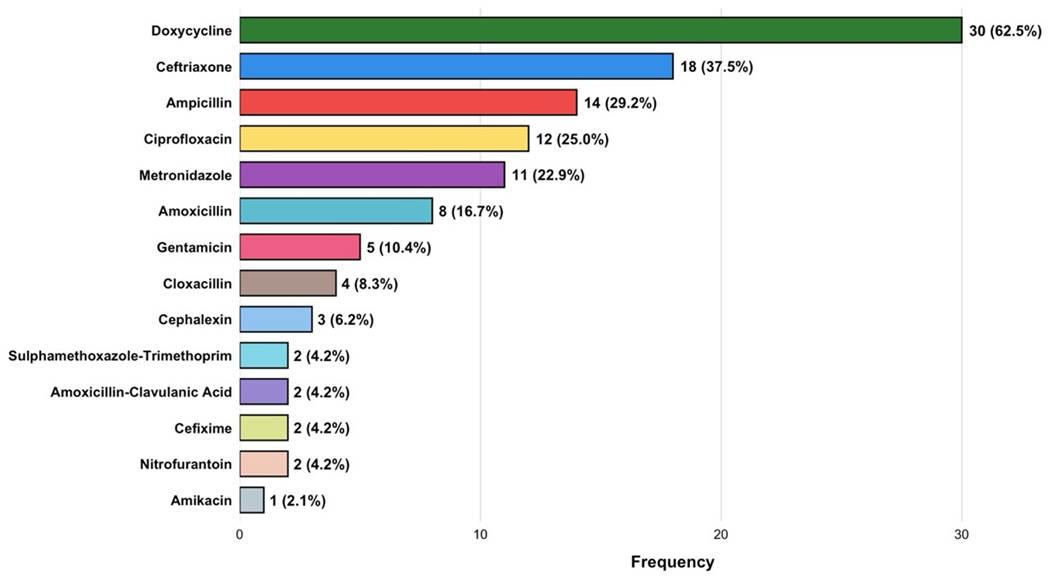
Figure 3: Distribution of previous antibiotic exposure across blood culture-positive episodes at the Eastern Regional Referral Hospital, Mongar, Bhutan, 2025
*Percentages are calculated based on the total episodes exposed (n = 48). Multiple antibiotic exposures were recorded per episode.
Distribution of multidrug-resistant organism and carbapenem-resistant organism
Of 79 blood culture isolates, 74 had ≥ 3 valid antimicrobial categories tested and were evaluable for MDRO classification (5 isolates were excluded as < 3 valid antimicrobial categories were evaluated; Pseudomonas aeruginosa, n=2; Streptococcus spp., n=3). Among evaluable isolates nearly one-fourth of the isolates were classified as MDRO (23.0 %, n = 17). For carbapenem resistance, 5 isolates were identified as CRO, representing 35.7% (5/14) of isolates tested against at least one carbapenem and 6.3% (5/79) of the total cohort. The highest proportion of the MDRO (47.1%) and CRO (80.0%) isolates were reported from Department of Medicine. E. coli was the most predominant organism in both MDRO (88.2%) and CRO (80.0 %). Other organisms contributed variably as presented in the Table 3.
Table 3: Distribution of Multidrug-resistant organisms and Carbapenem-resistant organisms by Department and Isolates among patients with Bloodstream Infections at the Eastern Regional Referral Hospital, Mongar, Bhutan, 2025.
|
Variable |
MDRO, n = 17 (%) |
CRO, n = 5 (%) |
|
|
Departments |
|||
|
|
Emergency |
4 (23.5) |
0 (0.0) |
|
|
Medicine |
8 (47.1) |
4 (80.0) |
|
|
Orthopedics |
2 (11.8) |
0 (0.0) |
|
|
Pediatrics |
0 (0.0) |
0 (0.0) |
|
|
Surgery |
3 (17.6) |
1 (20.0) |
|
Organisms |
|||
|
|
E. coli |
15 (88.2) |
4 (80.0) |
|
|
Pseudomonas aeruginosa |
0 (0) |
1 (20.0) |
|
|
Klebsiella pneumoniae |
2 (11.8) |
0 (0) |
|
|
Acinetobacter spp. |
0 (0) |
0 (0) |
|
|
Citrobacter sp. |
0 (0) |
0 (0) |
|
|
Enterobacter sp. |
0 (0) |
0 (0) |
|
|
Staphylococcus aureus |
0 (0) |
0 (0) |
|
|
Streptococcus spp. |
0 (0) |
0 (0) |
CRO: carbapenem-resistant organism; MDRO: multidrug- resistant organism
DISCUSSION
Given the rising burden of bloodstream infections alongside increasing antimicrobial resistance among common pathogens, particularly in LMICs, robust antimicrobial stewardship and evidence-based therapeutic strategies are essential. Effective implementation of such interventions requires a clear understanding of the local microbiological profile, baseline antimicrobial susceptibility pattern, and the burden of resistant organisms. Therefore, the study assessed the antimicrobial susceptibility pattern of isolated organisms and determined the proportion of MDRO among patients with positive blood cultures.
The present study observed a blood culture positivity rate of 8.1% after excluding coagulase-negative staphylococci, which is lower than several other regional studies such as 12.1% from Nepal and 28.26% from Pakistan.12,13 The relatively lower rate of positivity in the present study may be explained by the prior antibiotic exposure, which can suppress bacterial growth and culture yield.
The predominance of the gram-negative organisms with E. coli leading the isolates followed by Pseudomonas aeruginosa and dominance of Staphylococcus aureus among Gram-positive organisms aligns with reports from India, where a similar pattern has been described17. In contrast, studies from Nepal and some regions of India reported Gram-positive organism Staphylococcus aureus as the most commonly isolated organism 11,13 . The difference could be due to regional variation of prevalence of microorganisms causing bloodstream infection.
The antimicrobial susceptibility patterns observed in the current study can be interpreted through the lens of the WHO AWaRe classification, which categorizes antibiotics into Access, Watch and Reserve group based on the impact of different antibiotics and antibiotic classes on antimicrobial resistance 21. E. coli isolates demonstrated relatively high sensitivity to amikacin (86.7%), an Access group of aminoglycosides which is closely comparable to reports from Bangladesh and India 22,23. The high susceptibility to amikacin observed in our setting may be partly due to minimal prior exposure (Figure 3). Additionally, Amikacin requires prior authorization for its use as it is not available as an essential medicine in National Essential Medicine List, rendering it generally unavailable for routine use24. This restricted availability may potentially minimize broad spectrum selection pressure, though the direct impact of these health policy factors on local resistance patterns was not formally evaluated in this study. However, a significantly low susceptibility of E. coli to ampicillin (14.3%), also an Access group of antibiotics, is similar to the findings from Bangladesh and India reporting low susceptibility rates to commonly used access group antibiotic 22,23,25. This observed reduced susceptibility among Access group agents could hypothetically reflect broader community availability or historical usage patterns, highlighting a potential area for future longitudinal antimicrobial stewardship monitoring.
The high sensitivity of Pseudomonas aeruginosa to amikacin (100%) and gentamicin (88.9%), Access group of antibiotics and a high sensitivity to ciprofloxacin (94.4%), a Watch group of antibiotics in the present study appears more favorable than findings from India, where sensitivity was highest towards cefotaxime and colistin, a Watch and a Reserve group of antibiotics respectively25. The differences observed between the regions could hypothetically reflect geographic variations in antibiotic prescribing practice, and infection control and prevention measures. Moreover, the unregulated sale of antimicrobials over the counter without prescription could facilitate the emergence of resistance as reported across the globe26. However, these explanations remain speculative, as these factors were not evaluated in our study and warrant further comparative investigation.
The complete sensitivity of Staphylococcus aureus to cloxacillin (100%) an Access group of antibiotics, alongside high sensitivity to sulphamethoxazole-trimethoprim (85.7%), another Access group of antibiotics was observed in the present study, in contrast to regional studies reporting varying sensitivity rate12,13,23. From an AWaRe perspective, the observed susceptibility patterns among gram-positive isolates suggest that Access group antibiotics remain active in this small sample. While these findings align with the WHO AWaRe framework's emphasis on preserving Access agents as primary options where feasible, larger surveillance studies are needed before drawing definitive conclusions regarding empirical therapy guidelines.
MDROs and CROs represent one of the most pressing threats to global health security with a significant burden particularly in LMICs. In this study, 23.0% of the isolates were MDRO and 35.7% (5/14) of carbapenem-tested isolates were CRO [6.3% (5/79) across all isolates], with E. coli predominating in both categories (88.2% and 80.0% respectively). In contrast, neighboring regions have reported MDRO and CRO rates as high as 43% and 65% in 2021, respectively 13. The comparatively lower MDRO and CRO prevalence observed in the present study may hypothetically reflect the contextual factors specific to the region. Specifically, it is plausible that the predominantly public healthcare system and regulated antibiotic access with minimal over-the-counter availability by Bhutan Food and Drug Authority could contribute to this pattern 27. Furthermore, carbapenem use is limited as these agents are not included in the National Essential Medicine list and requires procurement through a named-patient medicine mechanism with prior authorization, limiting their use to facilities with culture and susceptibility testing capacity 24. This restriction policy could hypothetically explain the lower carbapenem resistance observed in our study. Despite the comparatively lower prevalence observed in the current study, it warrants proactive surveillance, strengthened infection prevention and control practices, and rational antibiotic use, due to rapid escalation of MDRO and CRO documented elsewhere in the region within relatively short timeframes.
All the Staphylococcus aureus isolates demonstrated complete susceptibility to cloxacillin indicating no methicillin-resistant Staphylococcus aureus strains in year 2025. The findings indicate that isolated staphylococcal strains remain fully susceptible to Access group of antibiotics, such as cloxacillin. This supports essential antimicrobial stewardship goals by optimizing patient clinical outcomes with safer first-line choices while minimizing the emergence of multidrug-resistant organisms.
This study has several limitations. The findings from a single-center tertiary care hospital study may not be generalizable to other regions in Bhutan. As this was a retrospective study, the sample size was determined by the availability of positive blood culture episodes during the study period, and all eligible cases were included rather than selecting a sample. The relatively low number of bloodstream infection episodes reflects the population size and healthcare setting of eastern Bhutan, as well as the possible prior antibiotic exposure which suppressed bacterial yield. Additionally, methodological factors may have introduced selection bias. First, the selective, tier-wise testing protocol meant that second-tier broad-spectrum antimicrobials were not universally tested across all isolates, resulting in smaller denominators and conditional susceptibility estimates reflecting incomplete testing panels rather than missing observational records; specifically, carbapenem susceptibility was tested on only a minority of isolates, meaning the CRO estimate rests on a small tested subset. Second, the potential retention of repeat isolates from individual patients may overrepresent specific resistant phenotypes. Third, the blanket exclusion of coagulase-negative staphylococci, while standard practice to avoid blood culture contamination, may have omitted clinically significant healthcare-associated-infections, means the overall positivity rate may not be directly comparable with studies that include or clinically adjudicate these organisms. Finally, the absence of molecular characterization of MDRO and CRO due to limitations in diagnostic facilities have limited the determination of the underlying mechanism of resistance.
Conclusion
Blood culture positivity rate in eastern Bhutan was 8.1% (excluding coagulase-negative staphylococci), with more than three-quarters of the isolates being Gram-negative organisms. E. coli was the predominant organism, with the highest sensitivity to amikacin. Nearly one-fourth of evaluable isolates were MDRO, while carbapenem resistance was identified in 5 isolates, representing 35.7% (5/14) of isolates tested against at least one carbapenem and 6.3% (5/79) of the total cohort. The study provides valuable baseline data to support local antimicrobial resistance surveillance. It also underscores the need for continuous microbiological surveillance, strengthened infection control and prevention practices and rational antibiotic use through effective antimicrobial stewardship to curb the emergence and spread of antimicrobial resistance.
DECLARATIONS
Ethics approval and consent to participate
Administrative clearance was obtained from the Ministry of Health, and waiver of ethical clearance was granted by the Institutional Review Board, Khesar Gyelpo University of Medical Sciences of Bhutan, with Ref. No. IRB/Waiver-Exempt/ PN/2025/064/114, dated 4th December 2025.
Availability of data and materials.
The datasets used and analyzed during the current study are available from the corresponding author upon reasonable request.
REFERENCES
1. Holmes CL, Albin OR, Mobley HLT, Bachman MA. Bloodstream infections: mechanisms of pathogenesis and opportunities for intervention. Nat Rev Microbiol. 2024 Apr 1;23(4):210. doi:10.1038/S41579-024-01105-2 PubMed PMID: 39420097.[PubMed][FullText][DOI]
2. Viscoli C. Bloodstream Infections: The peak of the iceberg. Virulence. 2016 Apr 2;7(3):248.doi:10.1080/21505594.2016.1152440 PubMed PMID: 26890622.[PubMed][FullText][DOI]
3. Lee Y, Kim YC, Seong J, Ahn S, Han M, Lee JA, et al. Predictors of bloodstream infection and its impact on mortality in septic arthritis: A 15-year review. Journal of Microbiology, Immunology and Infection. 2025 Jun 1;58(3):318-24. doi:10.1016/j.jmii.2025.02.006 PubMed PMID: 40059018.[PubMed][FullText][DOI]
4. Liu Y, Sun T, Cai Y, Zhai T, Huang L, Zhang Q, et al. Clinical characteristics and prognosis of pneumonia-related bloodstream infections in the intensive care unit: a single-center retrospective study. Front Public Health. 2023;11:1249695. doi:10.3389/FPUBH.2023.1249695 [PubMed][FullText][DOI]
5. Sepsis - NHS [Internet]. [cited 2026 Jun 8]. Available from: https://www.nhs.uk/conditions/sepsis/.[FullText]
6. Chen J, Huang H, Zhang R, Fu Y, Jing C. Risk factors associated with mortality and pathogen characteristics of bloodstream infection-induced severe sepsis in the pediatric intensive care unit: a retrospective cohort study. Front Cell Infect Microbiol. 2025 Feb 3;15:1492208. doi:10.3389/FCIMB.2025.1492208 PubMed PMID: 39963407.[PubMed][FullText][DOI]
7. Lamy B, Dargere S, Arendrup MC, Parienti JJ, Tattevin P. How to Optimize the Use of Blood Cultures for the Diagnosis of Bloodstream Infections? A State-of-the Art. Front Microbiol. 2016;7(MAY):697. doi:10.3389/FMICB.2016.00697 PubMed PMID: 27242721.[PubMed][FullText][DOI]
8. Fabre V, Sharara SL, Salinas AB, Carroll KC, Desai S, Cosgrove SE. Does This Patient Need Blood Cultures? A Scoping Review of Indications for Blood Cultures in Adult Nonneutropenic Inpatients. Clinical Infectious Diseases. 2020 Aug 22;71(5):1339-47. doi:10.1093/CID/CIAA039 PubMed PMID: 31942949.[PubMed][FullText][DOI]
9. Diekema DJ, Hsueh PR, Mendes RE, Pfaller MA, Rolston K V., Sader HS, et al. The Microbiology of Bloodstream Infection: 20-Year Trends from the SENTRY Antimicrobial Surveillance Program. Antimicrob Agents Chemother. 2019;63(7):e00355-19. doi:10.1128/AAC.00355-19 PubMed PMID: 31010862.[PubMed][FullText][DOI]
10. Kern W V., Rieg S. Burden of bacterial bloodstream infection-a brief update on epidemiology and significance of multidrug-resistant pathogens. Clinical Microbiology and Infection. 2020 Feb 1;26(2):151-7. doi:10.1016/J.CMI.2019.10.031 PubMed PMID: 31712069.[PubMed][FullText][DOI]
11. Gupta S, Shukla M, Meena M, Nag S, Islahi S, Singh S, et al. Bacteriological profile and antibiotic sensitivity pattern of bloodstream infections from a teaching institution in Eastern Uttar Pradesh, North India. J Family Med Prim Care. 2024 Sep;13(9):3871-8. doi:10.4103/JFMPC.JFMPC_1994_23.[PubMed][FullText][DOI]
12. Ejaz A, Khawaja A, Arshad F, Tauseef A, Ullah R, Ahmad I. Etiological Profile and Antimicrobial Patterns in Blood Culture Specimens in a Tertiary Care Setting. Cureus. 2020 Oct 17;12(10):e11000. doi:10.7759/CUREUS.11000 PubMed PMID: 33209556.[PubMed][FullText][DOI]
13. Khanal B, Shrestha LB, Sharma A, Siwakoti S. Bloodstream infections: trends in etiology and antimicrobial resistance in 10 years in Eastern Nepal. BMC Infect Dis. 2025 Dec 1;25(1). doi:10.1186/S12879-025-11413-4 PubMed PMID: 40781612.[PubMed][FullText][DOI]
14. Ahmed D, Nahid MA, Sami AB, Halim F, Akter N, Sadique T, et al. Bacterial etiology of bloodstream infections and antimicrobial resistance in Dhaka, Bangladesh, 2005-2014. Antimicrob Resist Infect Control. 2017 Jan 5;6(1). doi:10.1186/S13756-016-0162-Z PubMed PMID: 28070309.[PubMed][FullText][DOI]
15. Karajacob AS, Ibrahim F, Syed Omar SF, Zambry NS, Tay ST. Trends in bacterial and fungal bloodstream infections in Southeast Asia: A review of blood culture pathogens (2018-2024). Eur Rev Med Pharmacol Sci. 2025 Jul;29(7):356-74. doi:10.26355/EURREV_202507_37328 PubMed PMID: 40748345.[PubMed][FullText][DOI]
16. Global antibiotic resistance surveillance report 2025 WHO Global Antimicrobial Resistance and Use Surveillance System (GLASS) [Internet]. [cited 2026 Jun 17]. Available from: https://www.who.int/publications/i/item/9789240116337.[FullText]
17. Karuna T, Gupta A, Vyas A, Kumar S, Sampath A, Goel P, et al. Changing Trends in Antimicrobial Susceptibility Patterns of Bloodstream Infection (BSI) in Secondary Care Hospitals of India. Cureus. 2023 Apr 18;15(4). doi:10.7759/CUREUS.37800.[PubMed][FullText][DOI]
18. M100 | Performance Standards for Antimicrobial Susceptibility Testing [Internet]. [cited 2026 Jul 8]. Available from: https://clsi.org/shop/standards/m100/.[FullText]
19. Magiorakos AP, Srinivasan A, Carey RB, Carmeli Y, Falagas ME, Giske CG, et al. Multidrug-resistant, extensively drug-resistant and pandrug-resistant bacteria: An international expert proposal for interim standard definitions for acquired resistance. Clinical Microbiology and Infection. 2012;18(3):268-81. doi:10.1111/j.1469-0691.2011.03570.x PubMed PMID: 21793988.[PubMed][FullText][DOI]
20. Clinical and Laboratory Standards Institute. Performance Standards for Antimicrobial Susceptibility Testing [Internet]. 33rd ed. Vol. M100. Wayne, PA: Clinical and Laboratory Standards Institute; 2023 [cited 2026 Jul 9]. 152-161 p. Available from: https://iacld.com/UpFiles/Documents/672a1c7c-d4ad-404e-b10e-97c19e21cdce.pdf[FullText]
21. WHO. World Health Organization [Internet]. 2023 [cited 2026 Mar 27]. AWaRe classification of antibiotics for evaluation and monitoring of use, 2023. Available from: https://www.who.int/publications/i/item/WHO-MHP-HPS-EML-2023.04.[FullText]
22. Salsabil Ripa S, Sultana K, Mokarrama M. Antimicrobial Susceptibility Pattern in Blood Culture at Chattogram. Chattagram Maa-O-Shishu Hospital Medical College Journal. 2026 Feb 1;24(1):67-71. doi:10.3329/cmoshmcj.v24i1.82511[PubMed][FullText][DOI]
23. Yangzom T, Tsering DC, Kar S, Kapil J. Antimicrobial Susceptibility Trends among Pathogens Isolated from Blood: A 6-Year Retrospective Study from a Tertiary Care Hospital in East Sikkim, India. J Lab Physicians. 2020 Mar;12(1):3. doi:10.1055/S-0040-1712814 PubMed PMID: 32792787.[PubMed][Full Text][DOI]
24. National Essential Medicines List 2025 [Internet]. [cited 2026 Jul 8]. [Full Text]
25. Madavi V, Uike S, Aher C. ANTIBIOTIC SENSITIVITY PATTERN OF BLOOD CULTURE ISOLATES: A STUDY FROM A TERTIARY CARE HOSPITAL OF MAHARASHTRA. International Journal of Academic Medicine and Pharmacy. doi:10.47009/jamp.2024.6.2.217.[FullText][DOI]
26. Batista AD, Rodrigues DA, Figueiras A, Zapata-Cachafeiro M, Roque F, Herdeiro MT. Antibiotic Dispensation without a Prescription Worldwide: A Systematic Review. Antibiotics. 2020 Nov 1;9(11):786. doi:10.3390/ANTIBIOTICS9110786 PubMed PMID: 33171743.[PubMed][FullText][DOI]
27. Bhutan Food and Drug Authority - Royal Government of Bhutan [Internet]. [cited 2026 Jul 8]. Available from: https://bfda.gov.bt/[FullText]
|
AUTHORS CONTRIBUTION Following authors have made substantial contributions to the manuscript as under: ST: Conceptualization, literature search, data collection, data analysis, writing UR: data collection, review & editing manuscript. PW: review and edited final manuscript. CW: data collection and drafting manuscript. ND: data collection and drafting manuscript. UC: data collection and drafting manuscript. TW: review and edited manuscript. YD: data analysis, conducted literature research and reviewed final manuscript. Authors agree to be accountable for all respects of the work in ensuring that questions related to the accuracy and integrity of any part of the work are appropriately investigated and resolved. |
|
CONFLICT OF INTEREST None GRANT SUPPORT AND FINANCIAL DISCLOSURE None |