CASE REPORT
https://doi.org/10.47811/bhj.208
Primary gastric squamous cell carcinoma: a case report
Prabhat Pradhan1, 2, Jigme Wangchuk3, Ugyen Chophel4
1Department of Oncosurgery, Jigme Dorji Wangchuck National Referral Hospital, Thimphu, Bhutan
2Faculty of Postgraduate Medicine, Khesar Gyalpo University of Medical Sciences of Bhutan, Thimphu, Bhutan
3Department of Pathology, Jigme Dorji Wangchuck National Referral Hospital, Thimphu, Bhutan
4Department of General Practitioner, Wangdue Hospital, Wangdue Phodrang, Bhutan
Corresponding author:
Prabhat Pradhan
drprabhat0401@gmail.com
ABSTRACT
Primary gastric squamous cell carcinoma (SCC) is an extremely rare type of gastric cancer, accounting for less than 1% of all gastric malignancies. It has not previously been reported in Bhutan. We report a case of primary gastric squamous cell carcinoma in a 57-year-old Bhutanese woman, initially misdiagnosed as poorly differentiated adenocarcinoma. This report highlights the diagnostic challenges, management approach, and importance of histopathological accuracy.
Keywords: Gastric cancer; Squamous cell carcinoma
INTRODUCTION
Gastric cancer is the most common malignancy in Bhutan, with an incidence rate of 25.1 per 100,000 in men and 18.9 per 100,000 in women1. In the Bhutanese population, adenocarcinoma is the predominant histological type, accounting for 98% of gastric cancers2. Primary gastric SCC is a rare malignancy, representing less than 1% of all gastric cancers3,4. To date, no case of primary gastric SCC has been reported in Bhutan. Its atypical location and histological features often lead to diagnostic confusion with other malignancies or metastatic disease. There is no standardised treatment guideline, and the prognosis is generally poor.
We present the first case of primary gastric SCC in a 57-year-old woman in Bhutan.
CASE PRESENTATION
Patient Information and Clinical Findings
A 57-year-old woman presented with a two-month history of epigastric pain radiating to the back, early satiety, reduced appetite, occasional evening vomiting, and significant weight loss. She had a history of occasional consumption of locally brewed alcohol and chronic betel nut chewing, but no known comorbidities or past surgeries.
Physical examination, nutritional status, and performance scores were within normal limits (Eastern Cooperative Oncology Group [ECOG] performance status 1).
Diagnostic Evaluation and Treatment
An esophagogastroduodenoscopy (EGD) revealed a large, ulcero-proliferative, malignant-appearing ulcer in the antrum near the lesser curvature. Biopsy confirmed poorly differentiated adenocarcinoma. Notably, she had undergone a similar EGD two years earlier from the same site but failed to follow up; previous findings were unavailable. Recent endoscopic images were not available due to lack of recording facility in our institute. However, the endoscopy report did not mention any similar lesions in the oesophagus, gastroesophageal junction, or elsewhere in the stomach or duodenum.
Staging contrast-enhanced computed tomography (CECT) showed an enhancing wall thickening at the incisura (3 × 1.8 cm) near the pylorus, with minimal fat stranding around the left lobe of the liver. Multiple perigastric and peripancreatic lymph nodes were observed (largest 1.8 cm), but no ascites or distant metastases were noted-consistent with TNM (AJCC 8th edition) T3N2M0 staging. No lesions in the lungs, liver, oesophagus, or cervix were found.
She received three cycles of neoadjuvant FLOT (fluorouracil, leucovorin, oxaliplatin, and docetaxel) chemotherapy. This regimen was planned as part of a perioperative strategy, followed by radical distal D2 gastrectomy. Intraoperatively, a 3 × 3 cm nodular mass was found at the antrum, with a mucosal surface resembling a healed scar (Figure 1). No peritoneal, omental, pelvic, or hepatic involvement was seen.
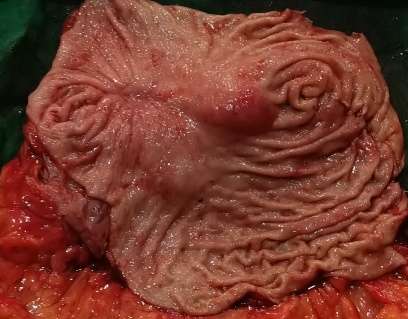
Figure 1. Nodular lesion with scarred mucosa and loss of folds observed in the resected stomach of a middle-aged woman at Jigme Dorji Wangchuck National Referral Hospital (JDWNRH), 2025.
Postoperative recovery was uneventful, and the patient was discharged on postoperative day six. Histopathology of the resected specimen revealed a moderately differentiated squamous cell carcinoma; no adenocarcinoma or adenosquamous components were identified. Initial slides were reviewed by onco-pathologists but immunohistochemistry (IHC) was not performed due to limited resources. Tumour cells infiltrated the subserosal layer, showing moderate eosinophilic cytoplasm, vesicular chromatin, prominent nucleoli, and keratin pearls (Figure 2). No tumour deposits were found in ten lymph nodes examined. There were no perineural or lymphovascular invasions. Treatment effect of neoadjuvant therapy was absent with extensive residual cancer and no evident tumour regression (poor to no response, score 3).
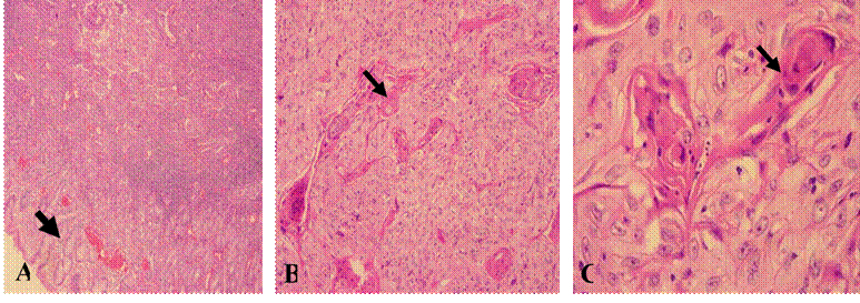
Figure 2. Microscopic images (haematoxylin and eosin, H&E-stained slides) of a resected stomach from a middle-aged woman at JDWNRH. (A) Low-power image showing nests of atypical squamous epithelial cells within the gastric wall; overlying normal gastric mucosa is noted (*40). (B and C) High-power images showing islands and nests of atypical squamous epithelial cells forming keratin pearls (*100 and *400).
Immunohistochemistry demonstrated positivity for P63 and CK5/6, and negativity for mucicarmine, confirming squamous cell carcinoma (Figure 3). Final staging was confirmed as ypT3N0.
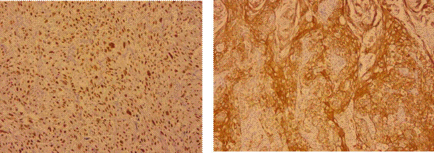
Figure 3. Immunohistochemistry of resected stomach from a middle-aged woman at JDWNRH, 2025, showing diffuse P63 nuclear and CK5/6 cytoplasmic staining of squamous cell carcinoma.
The discordance between the initial endoscopic biopsy showing poorly differentiated adenocarcinoma and the final surgical specimen demonstrating squamous cell carcinoma may be attributed to several factors. These include sampling error inherent to limited biopsy material, tumour heterogeneity with variable histological components, and the possibility of squamous metaplasia or squamous differentiation within a poorly differentiated tumour. Definitive diagnosis was established on the resected specimen following extensive histopathological evaluation, which remains the diagnostic gold standard.
Follow-up and Outcome
The patient was reviewed four weeks postoperatively and was doing well. In the absence of guidelines on the use of adjuvant chemotherapy for primary gastric SCC, and considering the final pathology (ypT3N0) indicating localised disease, a decision was made not to proceed with adjuvant chemotherapy.
The patient also declined further treatment, citing adverse experiences during prior chemotherapy. She was counselled regarding the risk of future recurrence, particularly given the limited lymph node harvest.
Subsequent follow-up six months after surgery-including EGD, CT scan, and CEA measurement-showed no evidence of recurrence (CEA 3.27). She was thereafter advised to follow up at her local hospital for convenience and with the oncologist as needed.
DISCUSSION
Primary gastric SCC is exceedingly rare, accounting for approximately 0.040.09% of all gastric malignancies4,5. Most reported cases originate from East and South Asia, particularly Japan, China, and Korea, with sporadic reports from other low- and middle-income countries6,7. Clinical presentation and imaging are indistinguishable from gastric adenocarcinoma, and preoperative misdiagnosis is common, as limited endoscopic biopsies may not capture the predominant squamous component4. The present case from Bhutan expands the limited literature from low-resource regions and reinforces the importance of diagnostic vigilance and histopathological-clinical correlation.
Table 1. Timeline of patient presentation and treatment.
|
Period |
Clinical course |
|
17th June 2024 |
Presented with epigastric pain radiating to the back, early satiety, reduced appetite, occasional evening vomiting, and significant weight loss. |
|
27th June 2024 |
Underwent EGD and biopsy from the lesion. |
|
15th July 2024 |
Reported as poorly differentiated adenocarcinoma. |
|
17th July 2024 |
Underwent CT of chest and abdomen: T3N2M0 |
|
19th July to 7th Sept 2024 |
Completed 3 cycles of FLOT. No major adverse events |
|
26th Sept 2024 |
CT response assessment after neoadjuvant chemotherapy (NACT)-no significant interval reduction |
|
28th Nov 2024 |
Radical D2 Distal Gastrectomy |
|
7th Jan 2025 |
HPE report: moderately differentiated squamous cell carcinoma (SCC). |
|
10th Jan 2025 |
Follow up with HPE report. No further treatment planned. |
|
2nd July 2025 |
Follow up EGD and CT showed no recurrence of disease. CEA 3.27 |
The diagnostic criteria established in 1967 by Parks8-(i) tumour not located at the cardia, (ii) no extension into the oesophagus, and (iii) absence of SCC elsewhere-remain relevant. In 2011, the Japanese Gastric Cancer Association added criteria requiring (i) presence of only squamous cell carcinoma cells and (ii) origin in the gastric mucosa9. Our case fulfilled all criteria for primary gastric squamous cell carcinoma.
The pathogenesis of primary gastric squamous cell carcinoma remains unclear. While it is more prevalent in males (male-to-female ratio approximately 6:1)10, our patient was female. Clinical presentation mimics that of gastric adenocarcinoma. Advanced primary gastric SCC generally carries a poorer prognosis compared to adenocarcinoma.
The etiological role of risk factors such as smoking, alcohol consumption, high-salt diets, obesity, and gastro-oesophageal reflux disease (GERD) is under investigation. Epstein-Barr virus (EBV) has been implicated in rare primary gastric SCC cases12,13.
There is no consensus on the treatment of primary gastric SCC. Surgery remains the mainstay for localised disease. Although some case series suggest limited benefit from neoadjuvant chemotherapy, 5-fluorouracilbased regimens may offer some response14. In our case, the FLOT regimen (fluorouracil, leucovorin, oxaliplatin, and docetaxel) did not yield significant tumour regression, but surgery proceeded successfully. The FLOT regimen was chosen considering the patient's relatively young age, good performance status, and its demonstrated survival benefit over other regimens for gastric adenocarcinoma, as reported in the FLOT4 trial14.
In low-resource settings, the diagnosis of primary gastric SCC is particularly challenging. Limited endoscopic biopsy samples may fail to represent the dominant histological component, leading to misclassification-especially in poorly differentiated tumours. Furthermore, lack of routine access to immunohistochemistry and subspecialty gastrointestinal pathology can contribute to diagnostic discordance. Close histopathological-clinical correlation, along with thorough examination of the resected specimen, remains essential for accurate diagnosis.
Reporting this case from Bhutan adds to the limited global literature and highlights the need for increased diagnostic vigilance and strengthened pathology services in resource-constrained healthcare systems.
CONCLUSION
This is the first reported case of primary gastric squamous cell carcinoma in Bhutan. The case highlights the need for diagnostic vigilance and close histopathological-clinical correlation to avoid mismanagement, particularly in resource-limited settings. Documentation of such rare malignancies is important for strengthening Bhutan's cancer registry and contributing to the global understanding of this uncommon disease.
ACKNOWLEDGEMENTS
We thank the patient and patient's attendants for consenting to the publication of clinical details and images. We thank the Pathology Department at JDWNRH for their assistance.
Ethical statement
Administrative approval was obtained from the Ministry of Health, Royal Government of Bhutan (date: 17/06/2025), and the hospital administration at the Jigme Dorji Wangchuck National Referral Hospital (JDWNRH).
Consent
Informed written consent was obtained from the patient in accordance with the consent process of the Institutional Review Board, Ministry of Health. No information related to patient identity or photographs that could lead to identification of the patient is presented.
REFERENCES
1. Tshomo U, Tshering P, Bagal S, Budukh A. Cancer Incidence and Mortality in Bhutan: 2019-2022. Ministry of Health, Bhutan, 2024.[Full Text]
2. Choden S, Wangmo C. The histopathological characteristics of gastric carcinoma in Bhutanese population. Bhutan Health Journal.[DOI]
3. Bonnheim DC, Sarac OK, Fett W. Primary squamous cell carcinoma of the stomach. Am J Gastroenterol. 1985;80:91-94.[PubMed]
4. Straus R, Heschel S, Fortmann DJ. Primary adenosquamous carcinoma of the stomach. Cancer. 1969;24:985-995.[DOI] [Full Text]
5. Muto M, Hasebe T, Muro K, et al. Primary squamous cell carcinoma of the stomach: a case report with a review of Japanese and Western literature. Hepatogastroenterology. 1999;46:3015-3018.[PubMed]
6. Wakabayashi H, et al. Primary squamous cell carcinoma of the stomach: a case report and review of the Japanese literature. J Gastroenterol. 1998;33:276-280.[PubMed]
7. Akce M, et al. Primary gastric squamous cell carcinoma: clinicopathologic features and outcomes from the SEER database. J Surg Oncol. 2019;120:873-879.[PubMed] [DOI] [FullText]
8. Parks RE. Squamous neoplasms of the stomach. AJR Am J Roentgenol. 1967;101:447-449.[Full Text]
9. Japanese Gastric Cancer Association. Japanese classification of gastric carcinoma: 3rd English edition. Gastric Cancer. 2011;14(2):101-112.[Full Text]
10. Chen Y, et al. Clinicopathological characteristics, treatment, and prognosis of 21 patients with primary gastric SCC. Gastroenterol Res Pract. 2016;2016:3062547.[PubMed] [DOI] [Full Text]
11. Meng Y, et al. Poorer prognosis in patients with advanced gastric SCC. Medicine (Baltimore). 2017;96:e9224.[PubMed] [DOI] [Full Text]
12. Katsura Y, et al. EBV-associated primary gastric SCC. Surg Case Rep. 2021;7:240.[PubMed] [DOI] [Full Text]
13. Takita J, et al. Immunohistochemical and molecular studies of primary gastric SCC. Hepatogastroenterology. 2005;52:969-974.[PubMed]
14. Al-Batran SE, Homann N, Pauligk C, et al. Perioperative chemotherapy with fluorouracil plus leucovorin, oxaliplatin, and docetaxel versus fluorouracil or capecitabine plus cisplatin and epirubicin for locally advanced, resectable gastric or gastro-oesophageal junction adenocarcinoma (FLOT4): a randomised, phase 2/3 trial. Lancet. 2019;393(10184):1948-1957.[PubMed] [DOI]
|
AUTHORS CONTRIBUTION Following authors have made substantial contributions to the manuscript as under: PP: Conceptualization, methodology, investigation, resources and writing-original draft JW: Conceptualization, methodology, investigation, resources and writing-original draft UC: Supervision, writing-review and editing Authors agree to be accountable for all respects of the work in ensuring that questions related to the accuracy and integrity of any part of the work are appropriately investigated and resolved. |
|
CONFLICT OF INTEREST |
|
None |
|
GRANT SUPPORT AND FINANCIAL DISCLOSURE |
|
None |